
5. Canine Parvovirus
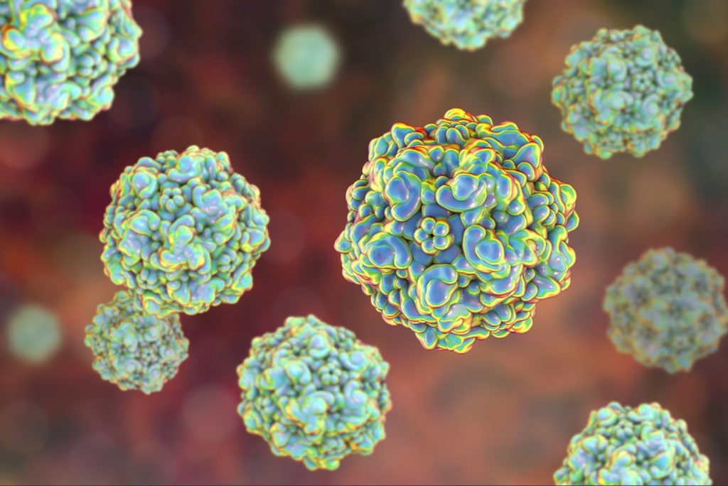
Canine parvovirus (CPV) is a highly contagious viral disease of dogs that commonly causes acute gastrointestinal illness in puppies. The disease most often strikes in pups between six and 20 weeks old, but older animals are sometimes also affected. A rare variant of the disease that may be seen in very young (neonatal) puppies is myocarditis (an inflammation of the heart muscle).
The virus that causes the disease known as “parvo”, canine parvovirus type 2 (CPV), first
emerged among dogs in Europe around 1976. By 1978 the virus had spread unchecked,
causing a worldwide epidemic of myocarditis and inflammation in the intestines
(gastroenteritis). We now know the virus is not limited to dogs but is capable of causing
infections in wild canines such as coyotes and wolves, and other wild animals, including
foxes, raccoons, and skunks. CPV is closely related to feline panleukopenia virus (FPV), a
virus that has been known since the 1920s to infect cats and mink, and other animals. CPV
probably arose as the result of 2 or 3 genetic mutations in FPV that allowed it to expand its
host range to infect dogs.
Three decades after its first appearance, CPV strikes puppies with the deadly disease much
less frequently due to the development of effective vaccines in the late 1970s, but outbreaks
still occur frequently and vaccinating your dog is of the utmost importance. Puppies and
adolescent dogs are especially susceptible to parvovirus, and you should avoid bringing your
puppy to public places where there is likely to be lots of viruses (animal shelters and kennels)
until after their vaccinations are complete.
Canine parvovirus can be found in almost any environment, but not every dog who comes
into contact with the virus becomes infected. Several factors come into play in infection,
including the immune status of the dog and the number of viruses the dog is exposed to. If the
combination of factors is just right and a dog does become infected, a specific sequence of
events is begun as the virus attacks the body.
Once a dog or puppy is infected, there is an incubation period of three to seven days before
the onset of the first symptoms. Inside the dog, CPV needs the help of rapidly dividing cells
to successfully cause disease, and the virus usually begins by attacking the tonsils or lymph
nodes of the throat. Once inside the lymph nodes, the virus typically invades lymphocytes (a
type of white blood cell) for one or two days, creating many copies of itself. These viruses
hitch a ride inside the lymphocytes, where they are sheltered from the host defenses, and
enter the bloodstream. Many of these CPV-infected lymphocytes are ultimately killed,
causing a reduction in the number of circulating lymphocytes, a condition called
lymphopenia.
Once in the bloodstream, the virus again targets rapidly dividing cells, hitting hardest in the
bone marrow and in the cells that line the walls of the small intestine. In very young dogs,
CPV can also infect the heart, leading to inflammation of heart muscle, poor function, and
arrhythmias.
In the bone marrow, the virus weakens the body’s ability to protect itself by destroying young
immune cells and causing a drop in the protective white blood cell count. This probably
makes it significantly easier for the virus to invade the gastrointestinal tract, where the virus
does its worst damage.
The virus causes this destruction by targeting the epithelium of the small intestine, the lining
that helps to absorb nutrients and provides a crucial barrier against fluid loss and bacterial
invasion from the gut into the body. The cells that make up the epithelial surface are short-
lived and are replaced continually by new cells born in the rapidly-dividing areas known as
the crypts of Lieberkühn. The virus invades these crypts where new epithelial cells are born
and disable the body’s ability to replenish the intestinal surface.
By preventing the replacement of old and dying cells with fresh new cells, the virus leaves
the intestinal surface unable to adequately absorb nutrients, prevent fluid loss into the stool,
or prevent bacteria from moving from the gut into the body. Severe diarrhea and nausea are
the initial results, but eventually, the intestinal surface can become so damaged that it begins
to break down, and the bacteria that are normally confined to the gut penetrate the intestine
walls and enter the bloodstream. This causes both significant fluid loss from diarrhea and
widespread infection inside the body. To make matters worse, the body’s immune system is
already weakened, as its ability to produce new white blood cells to combat infection has
been hampered by the invasion of CPV into the bone marrow. CPV is not always fatal, but
when it does kill, death is as a result of either dehydration and shock, along with the effects of
septic toxins produced by the intestinal bacteria roaming throughout the bloodstream.
Symptoms often associated with CPV include lethargy, depression, and loss or lack of
appetite, followed by a sudden onset of high fever, vomiting, and diarrhea. If your dog is
experiencing bouts of bloody diarrhea and/or vomiting, CPV is only one of several potential
culprits. Your veterinarian can run several tests to help determine whether your dog is
infected with CPV.
By far the most common and most convenient method of testing for the presence of CPV is
the fecal ELISA test. ELISA is an acronym for enzyme-linked immunosorbent assay, a
technology similar to that used in home pregnancy tests. In an ELISA test, antibodies to
parvovirus are immobilized on the surface of a testing chamber. A fecal sample is added to
the chamber, and antibodies attach to parvovirus proteins that may be present in the stool. A
color-changing chemical is then added to the chamber, and if parvoviruses have attached to
the antibodies, the chemical will change color and indicate a “positive” result. CPV fecal
ELISA tests can usually be completed by your veterinarian in less than 15 minutes. Though
the ELISA test is fairly accurate, it can occasionally produce false positive or false negative
results, so further testing may be necessary to confirm a diagnosis.
Veterinarians may also rely on a test that uses a technique called polymerase chain reaction
(PCR) to diagnose CPV from fecal samples. The CPV fecal PCR test detects small pieces of
viral DNA that are specific to CPV in the stool of an infected dog. This test is very accurate
(more so than CPV fecal ELISA), but requires that a fecal sample be sent to a laboratory that
specializes in performing PCR-based testing, so it generally requires more time than a CPV
fecal ELISA.
A simple measure of white blood cell count is often the clincher for a CPV diagnosis.
Because one of the first things the parvovirus infects is the bone marrow, a low white blood cell count can be suggestive of CPV infection. If a dog has both a positive ELISA reading
and a low white blood cell count, a fairly confident diagnosis of CPV may be made.
Treatment options for dogs suffering from CPV involve supportive care and management of
symptoms. Treatment options will vary, depending on how sick the dog is, but certain aspects
are considered vital for all patients.
A hospital stay is often necessary so that the dog can receive intravenous fluids and nutrients
to replace the vast quantities lost via vomiting and diarrhea. An intravenous drip is preferred
because the digestive tract of stricken dogs is usually in distress and can’t tolerate or absorb
what the dog needs. Blood transfusions may also be helpful to boost low blood cell counts
that may result from CPV infecting the bone marrow.
Antibiotics may be an appropriate therapy for a dog suffering from CPV, administered either
intravenously or as injections, to help fight the infection if intestinal bacteria have entered the
bloodstream. In addition, medications to control nausea and diarrhea are sometimes useful.
Many dogs will respond to medical therapy if it is initiated in a timely fashion, and those
dogs that recover from CPV infection retain lifelong protective immunity against the strain
that infected them.
Since the advent of several effective canine vaccinations for CPV, this infectious disease has
become much less of a threat to dogs. This does not mean, however, that CPV does not
remain a serious problem, and vaccination of your dog should not be considered an option –
it is a must.
Veterinarians usually administer the CPV vaccine as part of a combination shot which
includes, among others, the distemper, canine adenovirus, and parainfluenza vaccines. These
shots are given every 3 to 4 weeks from the time a puppy is 6 weeks old until he is at least 16
weeks of age. A booster vaccination is recommended one year later, and then at one at three-
year intervals thereafter.
The tiny parvovirus is extraordinarily hardy. They are capable of surviving for months
outside an animal, even through the winter, and are resistant to most household cleaning
products. Infected dogs can shed vast numbers of viruses, making it difficult to disinfect an
area once it has been exposed to an infected dog. These facts highlight the importance of
isolating any dog that is infected with CPV from other dogs. Given the fact that most
environments (including dog parks, lawns, and even homes) are not cleaned with disinfecting
products regularly, a puppy can be exposed to CPV without any warning, making vaccine
protection all the more important.
If your home and yard have been contaminated by an infected dog, there are steps you can
take to disinfect them before introducing a new dog or puppy. Despite its relative resistance
to cleaning agents, we do know that CPV can be inactivated by bleach. Cleaning with a
solution of one part bleach mixed with approximately 30 parts water is an acceptable method
for disinfecting any indoor area (including bedding, food/water bowls, and all surfaces) that
once housed an infected dog. There is evidence suggesting that CPV loses some of its ability
to infect an animal after one month in an indoor environment. Outside, you cannot (and
should not) bleach your lawn, but rain or watering can dilute the concentration of the virus
over time. This dilution, combined with the sanitizing effects of sunlight can bring the
number of viruses down to an acceptable level in a few weeks.